ALCTID - Annotated Lung CT Image Database
Introduction
Computed tomography (CT) of lungs provides a diagnostic tool for identifying a range of lesions and diseases visible in the obtained scans. For helping radiologists in a timely and efficient assessment of a large number of scans different machine learning methods have been applied for the detection and classification of abnormalities. However, before clinical usage of such algorithms it is necessary to achieve high accuracy of the algorithm. This is achieved through training and testing phases for which it is essential to have a database that would include a range of abnormalities in the lungs.
The ALCTID (Annotated Lung CT Image Database) database contains 2D lung CT images: 170 images annotated by an experienced thoracic radiologist and 170 images of healthy lungs.
Annotated abnormalities include cancerous tissue, enlarged lymph nodes, enlarged heart etc.
Annotations are presented as a rectangle over a ROI containing lesions. They are created and stored in the formats compatible with YOLO (Redmon et al., 2015} and Pascal VOC (http://host.robots.ox.ac.uk/pascal/VOC/). Annotations on 170 images have a total of 307 annotated ROIs.
If you use the database in your research please cite the following publication:
David Ivusic, Antun Petrak, Jelena Bozek, Sonja Grgic, "Annotated lung CT image database", 2022 International Symposium ELMAR, 2022, pp. 165-168, doi: 10.1109/ELMAR55880.2022.9899805
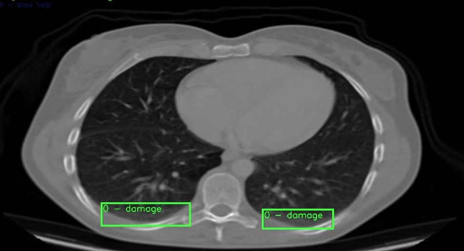
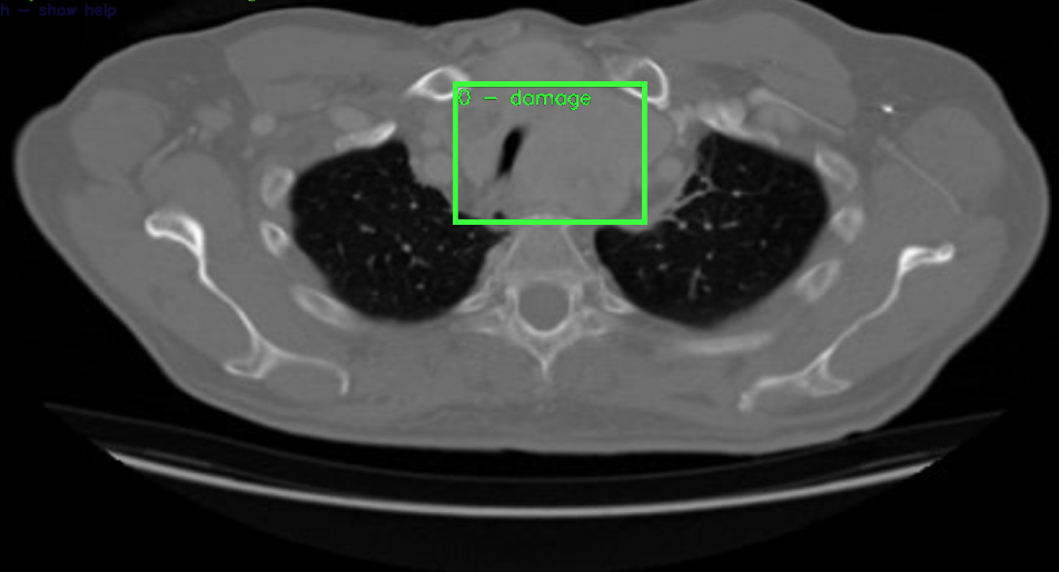
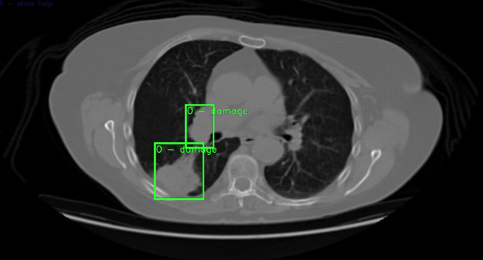
Example of annotations in the database:
 |
 |
 |
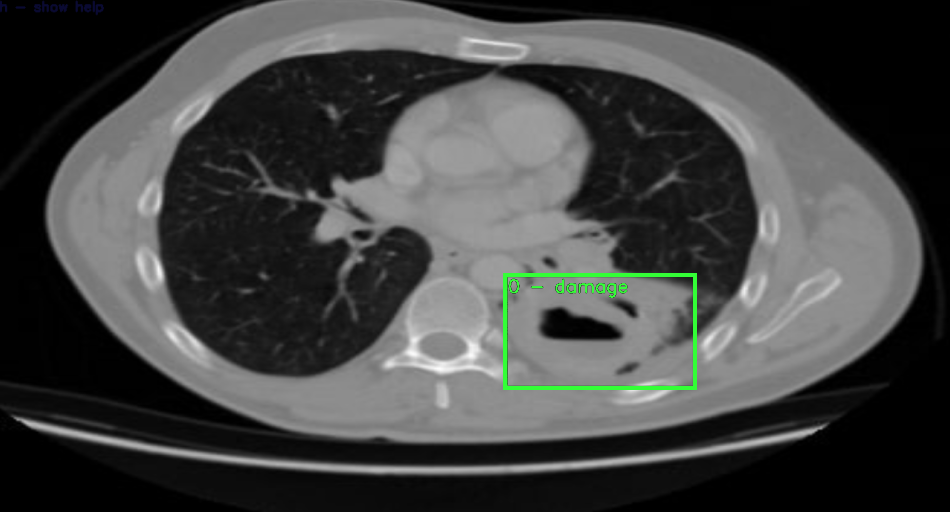 |
| An example annotation of an edema. | An example annotation of a large tumor in mediastinum. | An example annotations of a tumor and enlarged lymph nodes. | An example annotation of a possible tumor or lung abscess. |